Photonic crystals cause active color change in chameleons
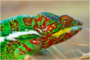
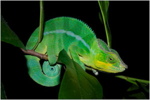
It is known for a long time that chameleons darken their skin through a process of melanin migration from the deep to the top layers of the dermis. When adult males are confronting a rival or a female, they rapidly switch between bright colors, changing from green to yellow, for example. It was believed that this variation was for the purpose of camouflage and that the mechanism of color change involves a shift of pigment, like in octopuses. Our study, recently published in Nature Communication, has provided evidence that this variation of color is due to the tuning of the lattice parameter of a three dimensional photonic structure made of a closed packed arrangement of submicron size guanine crystals in iridophore cells.


By Dirk van der Marel and Jérémie Teyssier, Department of Quantum Matter Physics, UNIGE
Based on article published in Nature Communication
Photometry analysis
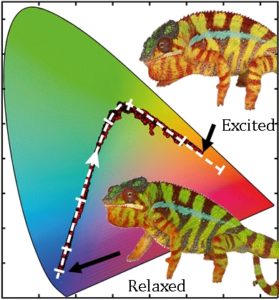
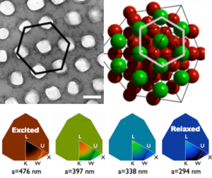
Color in chameleons is not only caused by pigments, “structural” colors (produced by interference of light on a periodic structure) plays also an important role. To provide evidence for the contribution of the photonic structure, we have developed a new method to extract tiny color variations on top of a constant background color from videos of chameleons while they are changing color. The figure below shows the first (“relaxed” chameleon) and last (“excited” chameleon) frames of a movie. The associated trace on the color chart shows a clear variation of photonic optical response across the entire spectral range from blue to orange.
Colorimetry at the single cell level
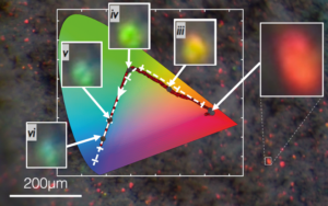
Applying osmotic pressure on skin samples allowed us to monitor the color evolution, at the level of a single iridophores cell, as the photonic crystal contracts. The associated color trace perfectly matches the in vivo experiment.
Photonic band structure
From crystal parameters measured using high quality transmission electron microscopy, we have modeled the band gap of the photonic structure using a plane wave expansion method.
Thus we derived the lattice dependence of the color, averaged over the entire Brillouin zone of the crystal. Experimental color traces recorded during in-vivo and ex-vivo experiments shown above, nicely follow the modeled lattice dependence of color (dashed white line on color charts)
This photonic structure represents a unique example of a soft, active, self-organized biologic structure. This opens many perspectives from a biological point of view, in particular to understand how such a perfect photonic structure develops and to investigate the mechanisms involved in the control of the geometry of iridophore cells. The optical efficiency of chameleon skin might also give birth to technological developments
A deep iridophore layer to prevent over-heating
In addition to the active top layer, we found a second, deeper layer of iridophores with larger and less organized guanine crystals. Using Fourier analysis on large size TEM images of skin cross sections, we shown that, spatial correlations are responsible for a strong enhancement of the reflectivity in the near infrared regions. This particular layer possessed by chameleons -and not by other species of lizards- could allow them a much better control of their energy balance, in particular when exposed to direct intense sunlight.
The United Living Color project
This project was initiated by Michel C. Milinkovitch (Laboratory of Artificial & Natural Evolution, University of Geneva), Dirk van der Marel (Quantum Materials Group, University of Geneva) and Matthias Zwicker (Computer Graphics Group, University of Bern) and funded by the Swiss National Foundation SINERGIA multidisciplinary program. In this project, we investigate the complex interactions between light and the vertebrate skin and how they generate a variety of astonishing phenomena of significant fundamental and applied interest.
UNIGE press release (in French)
UNIGE press release (in English)